igfold2d
IgFold2D
2D proteomaps for Ig-fold protein domains.
A small, dependency-free JavaScript widget that renders a folded-lace 2D map of an immunoglobulin domain, with residues placed at canonical positions on a grid indexed by the IgStrand universal numbering scheme (Tawfeeq et al., PLOS Comput Biol 2025).
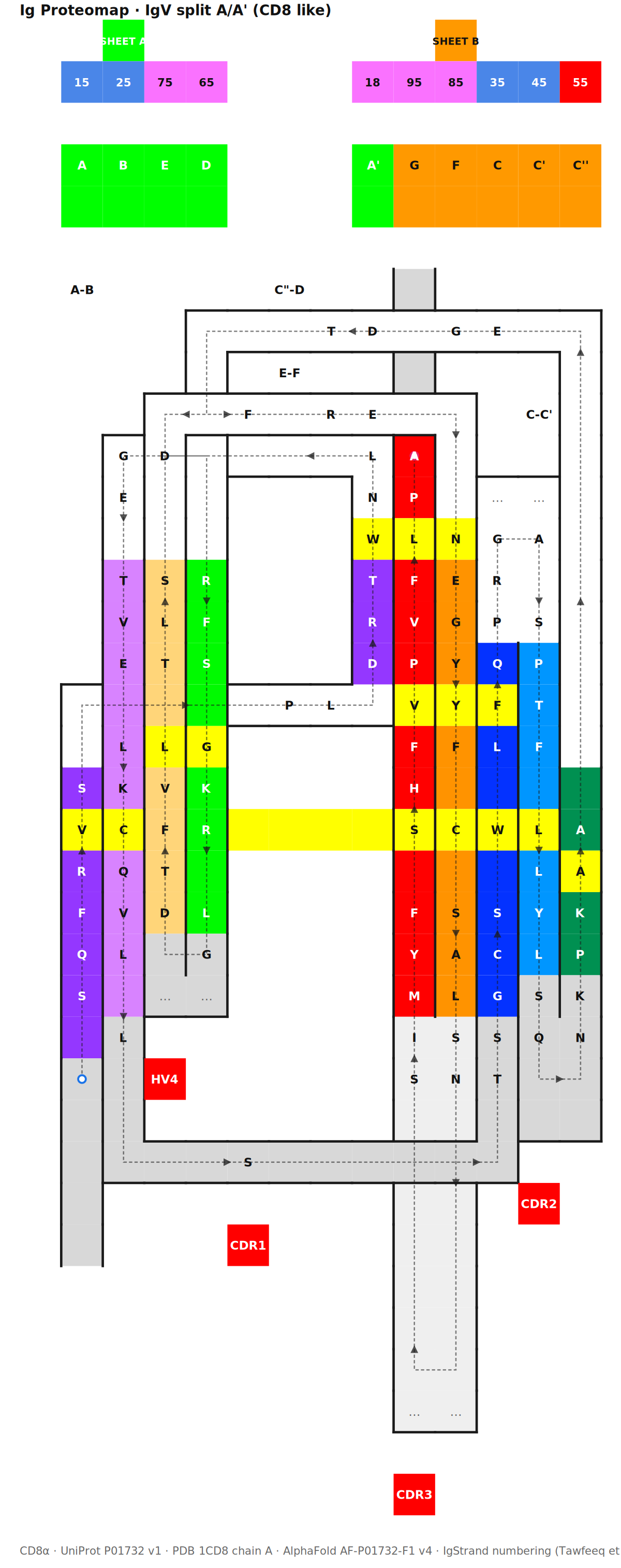
CD8α IgV domain. Each cell is addressable by Excel coordinate, IgStrand number, UniProt position, and PDB position. The dashed trace follows the chain N→C through anchor positions xx50 (yellow row).
Try it
- Live demo: vizomics.org/proteomaps — CD8α with bidirectional sequence ↔ proteomap sync
- Standalone HTML demo: clone the repo and open
examples/browser-widget/index.htmldirectly in a browser, no install - Static figure SVG: a pre-rendered, clickable, data-rich SVG is shipped with releases (
IgFold2D-CD8a.svg)
For iCn3D maintainers
👉 Start here: docs/icn3d-integration.md — three integration paths from “just add a link” to “full bidirectional 3D↔2D sync”.
The widget consumes the JSON output of iCn3D’s refnum.js (Ig domain detection + reference numbering) through a small adapter. No iCn3D internals are touched.
What it is
An Ig Proteomap is an explicit 2D projection of a 3D Ig-fold (β-sandwich) topology that preserves residue-residue contacts while making the sequential chain order visible.
Each cell of the 2D grid is multi-addressable:
- Excel coordinate (e.g.
L10) (row, col)pair- IgStrand number (e.g.
2550for the B-strand anchor) - UniProt / PDB / AlphaFold position
The widget supports:
- 5 display modes (residue letter, IgStrand #, UniProt #, PDB #, Excel address)
- Click and hover interactivity with a complete cell info panel
- Dashed chain trace following the IgStrand sequence A → G with direction chevrons
- Bidirectional highlight between linear sequence view and 2D proteomap
- Extensible feature vector per cell (conservation, contacts, SASA, mutations, …)
Quick start
Browser, drop-in
<div id="map"></div>
<script src="src/igfold2d.js"></script>
<script>
  Promise.all([
    fetch('templates/IgV-split-AAprime.json').then(r => r.json()),
    fetch('assignments/CD8a-P01732.json').then(r => r.json())
  ]).then(([template, assignment]) => {
    IgFold2D.render({
      container: '#map',
      template,
      assignment,
      sources: {
        uniprot:   { id: 'P01732', version: 1 },
        pdb:       { id: '1CD8', chain: 'A' },
        alphafold: { id: 'AF-P01732-F1', version: 4 }
      }
    });
  });
</script>
NPM (planned)
npm install igfold2d
import IgFold2D from 'igfold2d';
Repository layout
src/
  igfold2d.js              Widget core (~430 lines, ~13 KB minified, no deps)
templates/                 JSON template files (one per Ig variant)
  IgV-split-AAprime.json   IgV split A/A' (CD8-like) — 9 strands
assignments/               Pre-computed residue → IgStrand mappings
  CD8a-P01732.json         CD8α IgV domain (107 residues, hand-curated)
examples/
  browser-widget/          Standalone HTML demo (no server, no install)
website/                   The vizomics.org/proteomaps page
docs/
  icn3d-integration.md     ← How to integrate into iCn3D
  topology-rules.md        Fold construction rules
  igstrand-spec.md         IgStrand numbering reference
  api.md                   Widget API reference
The folding rules (short version)
Full reference in docs/topology-rules.md. Highlights:
| Rule | Description |
|---|---|
| No crossing | The lace never visually crosses itself on colored cells |
| Turns at strand ends | 90° turns happen above or below strands, never mid-loop |
| Minimize bends | Each strand-to-strand transition uses the minimum number of right-angle turns |
| Anchor alignment | All xx50 positions sit on one horizontal line |
| Passes-behind | Where a 3D chain crosses behind another, the 2D trace may visibly cross (dashed) or break (gap) |
| Placeholder traversal | Empty cells can be traversed as bridges; future residue insertions may land there |
IgStrand numbering
4-digit codes ijxx:
i(thousands): strand id — A=1, B=2, C=3, C’=4, C”=5, D=6, E=7, F=8, G=9j(hundreds): 5 for canonical strands; 8 for A’ (split case)xx: position centered on 50 (the anchor)- Insertions: A−=14xx, A++=13xx, A+=16xx, G+=96xx, etc.
Full spec in docs/igstrand-spec.md.
Status & roadmap
| Version | Status | Scope |
|---|---|---|
| v0.1 | ✅ shipped | Hand-curated CD8α template, browser widget, sequence↔map sync |
| v0.2 | 🚧 planned | More templates: IgC1, IgC2, IgI, FN3 |
| v0.3 | 🚧 planned | Live IgStrand assignment via iCn3D refnum.js |
| v0.4 | 🚧 planned | Feature overlays: conservation, contacts, SASA, mutations |
| v0.5 | 🚧 planned | Multi-domain view with inter-domain contacts |
License
MIT — see LICENSE.
Citation
If you use IgFold2D in your research, please cite the underlying numbering scheme:
Tawfeeq M, et al. (2025) A universal residue numbering scheme for the Immunoglobulin-fold (Ig-fold). PLOS Comput Biol 21(4): e1012813. doi:10.1371/journal.pcbi.1012813
Acknowledgments
Developed by Philippe Youkharibache and contributors at vizomics.org. The lace-folding concept extends prior work by the ISMB-2021 hackathon team (Manfredi, Patel, Cachau, Byrum) — see hackathonismb/2D-Maps-for-Ig-domains-and-for-RBDs. iCn3D integration consultation: NCBI Structure team.